AI Engineering · Computer Vision
PUBLISHED RESEARCHMalaria Parasite Stage Detection
YOLOv8 and Faster R-CNN ensembles with LAB color-space augmentation, exported to ONNX for lightweight deployment in diagnostic workflows.
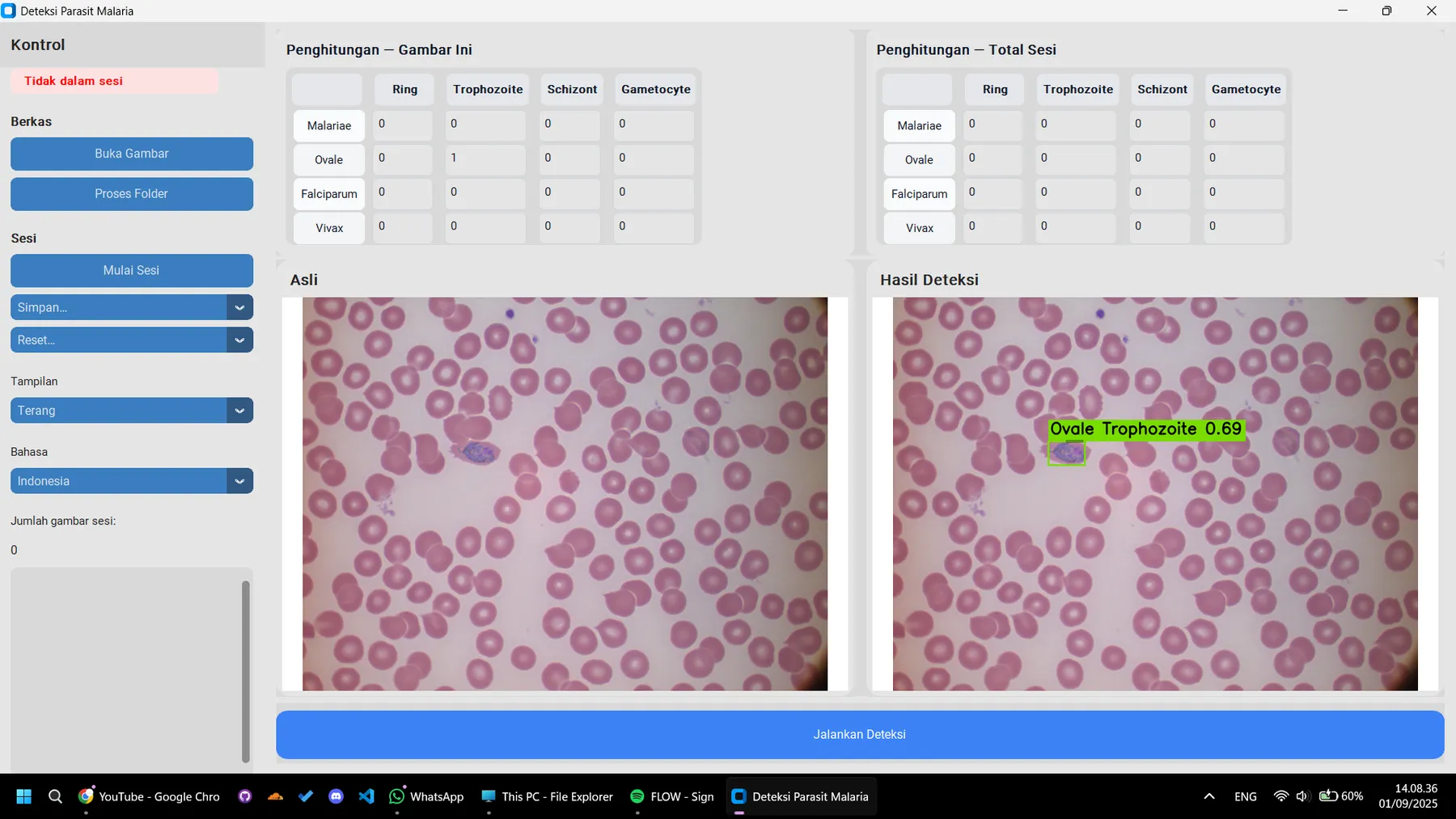 FIG // 02.A
mAP50 improvement (0.61 → 0.69) via LAB color-space feature insertion
Plasmodium species and life stages detected
Problem
Manual microscopy for malaria diagnosis is slow, requires trained lab technicians, and results vary between readers. Clinics without a resident parasitologist often wait days for a confirmed species and life-stage read — time that matters for treatment decisions. The goal of this thesis project was to build an assistive detection tool accurate enough to support (not replace) a lab technician’s read, while staying light enough to run on the hardware most labs already have.
Approach
The core challenge was distinguishing Plasmodium falciparum, vivax, malariae, and ovale across their different life stages inside stained blood smear images — a fine-grained detection problem where species look nearly identical under standard RGB preprocessing.
- Trained and benchmarked YOLOv8 and Faster R-CNN as parallel candidates, comparing precision/recall trade-offs across species classes.
- Introduced a LAB color-space feature insertion step ahead of the detection backbone, which made subtle staining differences between species more separable than in raw RGB.
- Iterated on augmentation (rotation, color jitter, mosaic) specifically tuned for microscopy imagery rather than natural-image defaults.
Architecture
The trained ensemble is exported to ONNX so inference doesn’t depend on a Python environment or GPU. The final tool packages into a standalone executable, so a lab can drop it onto a workstation without installing PyTorch, CUDA, or any Python tooling at all — a deliberate choice given the actual deployment environment is a clinical lab, not a data science workstation.
Result
The LAB color-space feature insertion lifted mAP50 from 0.61 to 0.69, a meaningful jump for a fine-grained, four-class detection task. The work was presented at AMLDS 2025 in Tokyo and published in IEEE proceedings, with the underlying methodology now feeding into further research-assistant work on medical image enhancement.